Service Categories
BIOINFORMATICS
ROSALIND - Fast Genomics Data Analysis - OnRamp
Service:
Check the Video to see How it Works
Capability:
The ROSALIND™ platform provides a biologist-friendly experience with instant results to visualize, interpret and discover more in your data. ROSALIND™ offers researchers, biologists and life scientists' solutions to analyze their own data without needing prior bioinformatics experience. ROSALIND is intended to speed up and simplify the process of genomic data analysis from experiment setup and QC to interactive data visualization and interpretation.
Experiment Types:
NanoString assays, scRNA-seq, Total RNA-seq ChiP-seq, and Small RNA-seq.
Pricing:
Inside rate:
$34 per unit
Outside rate:
For other assays, please submit a request at e6molina@ucsd.edu
Custom quote - Contact e6molina@ucsd.edu
Please inquire with Dr. Elsa Molina for more information.
Digital Spatial Profiling
GeoMx Digital Spatial Profiler - nanoString
Instrument:
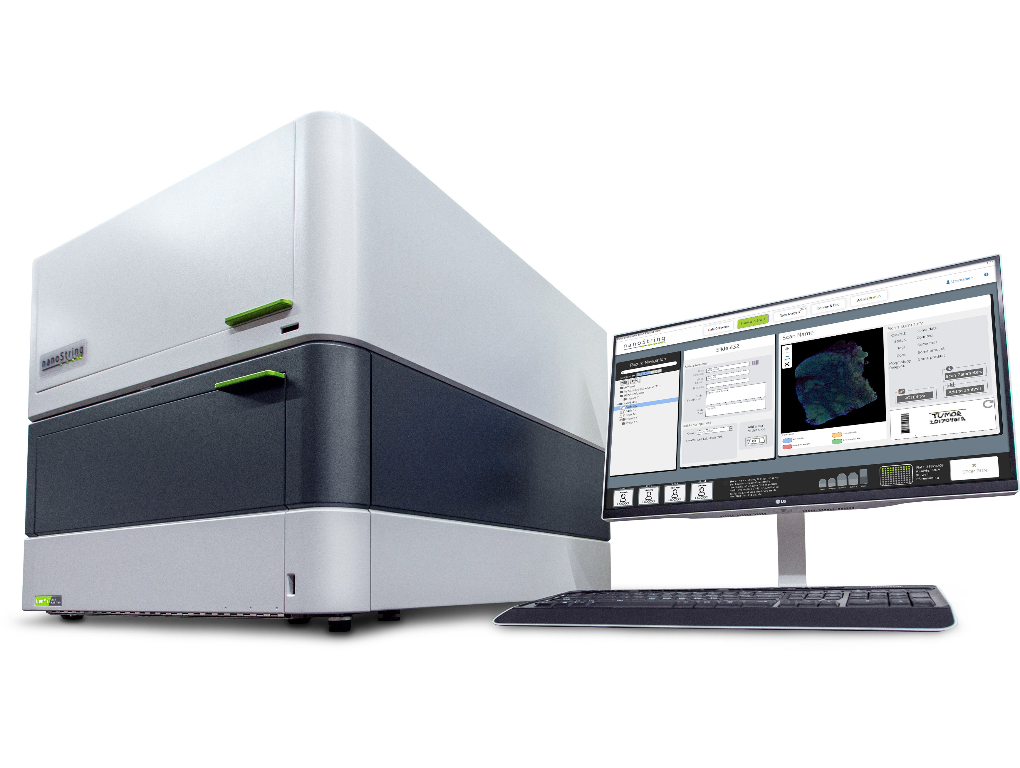
GeoMx Digital Spatial Profiler
Core Facility: Stem Cell Genomics Core
ELECTROPORATION
Amaxa Nucleofector
Instrument:
Schedule Type:
Equipment
Capability:
The Nucleofector Device is an apparatus which delivers unique electrical parameters. The electrical parameters are different from any other commercially available electroporation device and are especially developed to facilitate DNA transport into the cell nucleus. Each electrical setting is displayed as a distinct program which has been adapted to the requirements of a particular cell type.
Core Facility: Human Embryonic Stem Cell Core
FLOW CYTOMETRY
BDInflux Sorter
Instrument:
Schedule Type:
Flow Cytometry
Capability:
The BD Influx fluidics system features a unique acoustical coupling in the nozzle assembly to reliably create droplets for sorting, while ensuring low shear stress to optimize cell viability, even at high pressures. To support aseptic sorting, the fluidics path is easily removed and replaced with the optional disposable fluidics kit.
Core Facility: Human Embryonic Stem Cell Core
Aria Fusion Sorter
Instrument:
The fusion of safety, performance, and sorting
Capability:
The BD FACSAria™ Fusion flow cytometer delivers exceptional multicolor performance and ease-of-use. First introduced as the BD FACSAria™ sorter in 2003, this sorting knowhow is combined with best-in-class biosafety expertise for a comprehensive advanced cell sorter and biosafety solution. The fully integrated biosafety cabinet meets emerging operator and sample protection requirements as well as global standards for bioprotection.
Core Facility: Human Embryonic Stem Cell Core
BD LSR Fortessa
Instrument:
Schedule Type:
Flow Cytometry
Capability:
The BD LSRFortessa can be ordered with up to four lasers—blue, red, violet, and UV—which provides a flexibility in laser wavelengths so assay design can be optimized using the latest fluorescent dyes and substrates. The instrument can accommodate the detection of up to 18 colors simultaneously with a defined set of optical filters that meet or exceed the majority of today’s assay requirement.
Core Facility: Human Embryonic Stem Cell Core
BD LSRFortessa X-20
Instrument:
BD LSRFortessa™ X-20
Designed for limited space and boundless potential
The BD LSRFortessa™ X-20 cell analyzer delivers high-performance multicolor analysis with the most compact footprint in its class at just 30" x 29" (76.2 x 73.7 cm) and a height of 30" (76.2 cm).
The analyzer can be configured with up to 5 lasers to detect up to 20 parameters simultaneously to support ever increasing demands in multicolor flow cytometry. A wide range of up to 34 laser choices is available as excitation sources, including blue, red, violet, yellow-green, and UV
Contact us today to learn more about trading in your BD TM LSR II system and take advantage of our trade-in programs.
The BD LSRFortessa™ X-20 fits most flow cytometry analyzer needs, and can be ordered with up to 5 lasers-most commonly, blue, red, violet, yellow-green, and UV-which provides flexibility in laser wavelengths. This allows researchers to optimize assay design by selecting the latest fluorescent dyes and antibodies in experimental protocols. The instrument can accommodate the detection of up to 18 colors simultaneously with a defined set of optical filters that meet today's assay requirements.
The BD LSRFortessa X-20 is designed to support your current assay requirements. In addition, as future needs arise, optical capability can be added or upgraded.
A compact footprint and height of 76.2 x 73.7 x 76.2 cm (30 x 29 x 30 in.) allows the BD LSRFortessa X-20 to fit easily on the benchtop for the cost-effective use of valuable laboratory space. In addition to the reduced size, design enhancements provide easier access to bandpass filters and mirrors, simplifying changes in the instrument configuration.To improve experimental workflow, the optional BD TM High Throughput Sampler (HTS) provides rapid, fully automated sample acquisition from microtiter plates. In high-throughput mode, the HTS option can speed through a 96-well plate in less than 15 minutes with less than 0.5% sample carryover from one well to the next. Low carryover is essential to ensure data quality in research applications.
Core Facility: Human Embryonic Stem Cell Core
BD ARIA ii Sorter
INSTRUMENT:
Meet the new BD FACSAria II high-speed, fixed-alignment cuvette cell sorter.
Capability:
The first generation BD FACSAria™ system brought the complex world of cell sorting to a broader audience of researchers. Now the new BD FACSAria II system makes cell sorting even easier and more accessible.
Designed with input from customers around the world, the new BD FACSAria II improves ease of use, flexibility, and aseptic capability to make it simply the best choice for consistent results in sorting across a broad range of applications.
The BD FACSAria II incorporates the unique fixed alignment technology that revolutionized cell sorting over five years ago with the introduction of the BD FACSAria. Fixed alignment minimizes startup time and improves reproducibility. Gel coupling to the objective lens improves collection efficiency and increases sensitivity and resolution needed for multicolor sorting applications.
Core Facility: Human Embryonic Stem Cell Core
GENE EXPRESSION
nCounter® MAX Analysis System
Instrument:
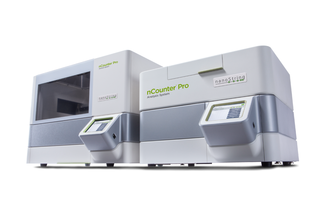
nCounter® MAX Analysis System
Fully automated and easy-to-use, the nCounter® MAX Analysis System provides everything you need to cost-effectively complete your projects in record time. More accurate and highly multiplexed than qPCR and simpler than NGS, the nCounter® MAX allows for digital examination of multiple pathways in a single tube in an extraction-free workflow requiring only 15 minutes of hands on time. Accelerate your research by spending less time on sample prep and perform your own data analysis using the included nSolver Analysis Software.
- Boost Productivity: Intuitive workflow with only fifteen minutes of hands-on time from sample to data. Separate Digital Analyzer and Prep Station units help eliminate bottlenecks in sample processing and data collection.
- Simplify Analysis: No need for a specialized Bioinformaticist. Results generated as direct counts and reported in a standard CSV file that can be imported into your favorite application or use the included nSolver Analysis Software for convenient data analysis.
- Detect Small Fold Changes: Eliminate cDNA synthesis, amplification, and library prep so you experience less technical variation in your assay and reduce the need for experimental replicates.
- Expandable Throughput: Add a second Prep Station when needed and double your capacity to match a growing number of users. This configuration enables processing of up to 96 lanes per day and 384 samples with sample plexing.
- Secure Access: An enterprise software package is included for laboratories that require enhanced security. Control user access, automate data flow, and generate audit logs.
- Unattended Data Collection: The Digital Analyzer can load up to six cartridges at a time for unattended scanning overnight. Random access enables users to pause data collection to load or remove additional cartridges without waiting for the run to finish.
*Inside users include UC San Diego, Salk Institute for Biological Studies, Sanford Burnham Prebys Medical Discovery Institute, and La Jolla Institute for Immunology.
Resources:
Stem Cell Characterization Panel
Note: Some consumables are also available for purchase. Please inquire with Dr. Elsa Molina for more information.
Core Facility: Stem Cell Genomics Core
MOLECULAR IMAGING INSTRUMENTS
11.7 T horizontal Bruker BioSpec MRI
Instrument
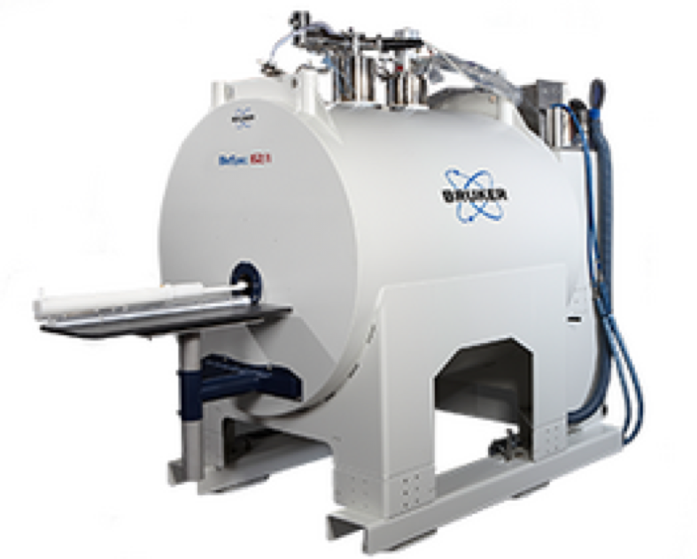
11.7 T horizontal Bruker Biospec MRI.
Capabilities- 16 cm room temperature bore
- Gradients with strength 750 mT/m, 90 mm clear-bore access
- Laser guided motorized positioning system
- Beds and stereotaxic hardware
- Ambient temperature control and body temperature monitoring
- Monitoring system for ECG and respiration triggering acquisitions
- Multiple Bruker RF coils:
- Circularly polarized 1H volume coil with inner diameter 38 mm (T12247V3)
- Circularly polarized 1H volume coil with inner diameter 72 mm (T11232V3)
- Body volume coil (T11440V3)
- Surface array coil (T11766V3)
- Surface array coil (T11473V3)
- Double-tuned 1H/19F volume coil (T12800V3)
- Double-tuned 1H/19F volume coil (RAPID Biomedical)
- Double-tuned 1H/19F surface coil (T11620V3)
- Double-tuned 1H/31P surface coil (T11619V3)
- Double-tuned 1H/13C surface coil (T11618V3)
Core Facility: MICS
Bioluminescense/flourescence Perkin Elmer IVIS Spectrum System with 3D imaging and spectral unmixing
Instrument
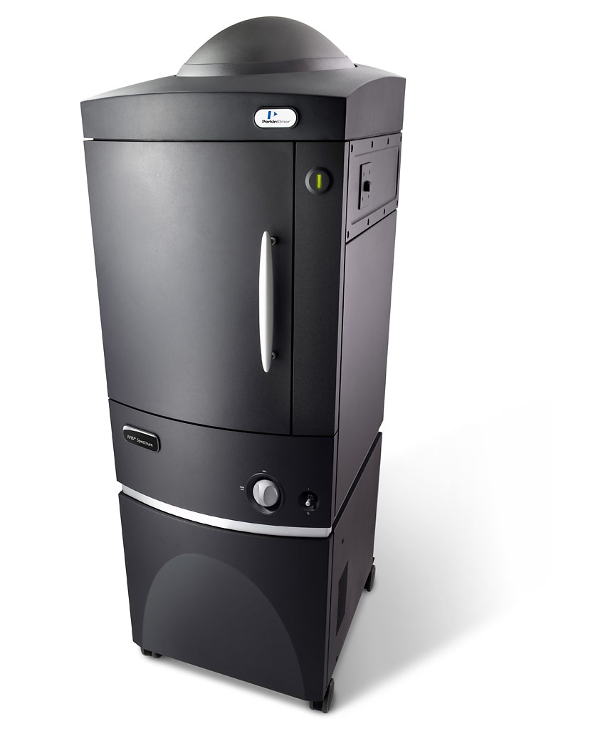
Perkin Elmer IVIS Spectrum system
CapabilitiesThe IVIS® Spectrum in vivo imaging system combines 2D optical and 3D optical tomography in one platform. The system uses leading optical technology for preclinical imaging research and development ideal for non-invasive longitudinal monitoring of disease progression, cell trafficking and gene expression patterns in living animals.
An optimized set of high efficiency filters and spectral un-mixing algorithms lets you take full advantage of bioluminescent and fluorescent reporters across the blue to near infrared wavelength region. It also offers single-view 3D tomography for both fluorescent and bioluminescent reporters that can be analyzed in an anatomical context using our Digital Mouse Atlas or registered with our multimodality module to other tomographic technologies such as MR, CT or PET.
For advanced fluorescence pre-clinical imaging, the IVIS Spectrum has the capability to use either trans-illumination (from the bottom) or epi-illumination (from the top) to illuminate in vivo fluorescent sources. 3D diffuse fluorescence tomography can be performed to determine source localization and concentration using the combination of structured light and trans illumination fluorescent images. The instrument is equipped with 10 narrow band excitation filters (30nm bandwidth) and 18 narrow band emission filters (20nm bandwidth) that assist in significantly reducing autofluorescence by the spectral scanning of filters and the use of spectral unmixing algorithms. In addition, the spectral unmixing tools allow the researcher to separate signals from multiple fluorescent reporters within the same animal.
Features & Benefits
- High-sensitivity in vivo imaging of fluorescence and bioluminescence
- High throughput (5 mice) with 23 cm field of view
- High resolution (to 20 microns) with 3.9 cm field of view
- Twenty eight high efficiency filters spanning 430 – 850 nm
- Supports spectral unmixing applications
- Ideal for distinguishing multiple bioluminescent and fluorescent reporters
- Optical switch in the fluorescence illumination path allows reflection-mode or transmission-mode illumination
- 3D diffuse tomographic reconstruction for both fluorescence and bioluminescence
- Ability import and automatically co-register CT or MRI images yielding a functional and anatomical context for your scientific data.
- NIST traceable absolute calibrations
- Gas anesthesia inlet and outlet ports
- Class I Laser Product
Core Facility: MICS
PET-SPECT-CT Integrated Trimodal Siemens Inveon Preclinical System
Instrument
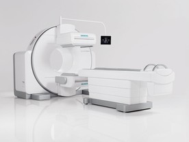
Capabilities
Visit here for more information on the Inveon PET-SPECT-CT Siemens trimodal system.
Core Facility: MICS
Ultrasound Visualsonics Vevo 2100
Instrument
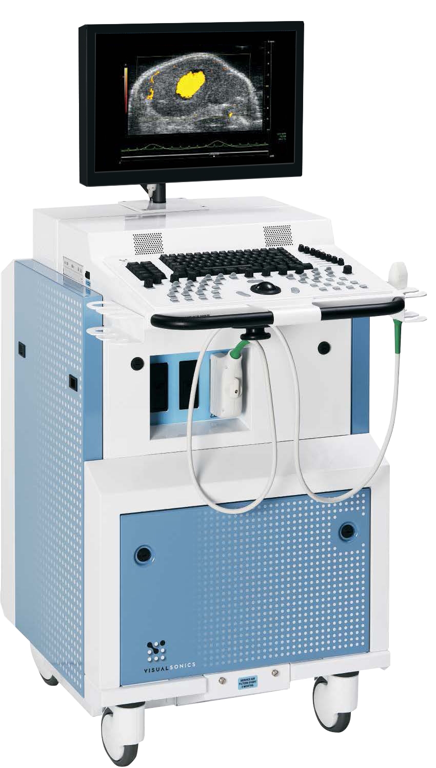
The Vevo® 2100 system expands the functionality, flexibility and image quality of the earlier Vevo 770 system
Capabilities
The Vevo 2100 operates at frequencies never before achieved with solid-state array transducers.
The new MicroScan™ transducers provide increased frame rates, superb contrast, unrivaled resolution and a wider field of view.
Features Include:
- Superior resolution and image uniformity through entire field of view
- 30 micron resolution
- Frame rates up to 740 fps
Expandable Imaging and Processing Options
- Color and Power Doppler Modes for blood flow quantification
- M-Mode single line acquisition for high-temporal resolution in LV functional analysis
- 3D-Mode Imaging & Volume Analysis
- Nonlinear Contrast Imaging
- VevoStrain™ Analysis software for cardiac research
- Advanced measurements & quantification
Software analysis and data management:
Key Features
- Ideal for long term studies with ability to add images/data to an existing study
- View images simultaneously for consistency and comparison
- Reserve time on the system - perform data analysis on an off-line computer
Data Management and Export
Analyze, label, annotate and archive images and studies through a flexible, open-architecture system.
- Export a list of acquired studies/series/images to paste into lab book
- Easily export with and without annotations/measurements (without duplication of data)
- Easily export with and without physiological parameters (without duplication of data)
Export data as TIFF, BMP, AVI uncompressed AVI, MS Video9 and DICOM
Core Facility: MICS
MICROSCOPY
Confocal Laser Scanning Biological Microscope FV1000 FluoView with Two-photon – Olympus
Instrument:
Schedule Type:
Microscopes
Capability:
This model features the world's first twin scan system. This system enables time-resolved spectroscopy of living intercellular contents and also makes it possible to analyze objects down to a 2nm wavelength resolution.
Core Facility: Stem Cell Genomics Core
Confocal Microscope LSM 780 – Zeiss
Instrument:
Zeiss LSM 780 Confocal Microscope
Capability:
The PTC laser concept of LSM 780 is revolutionary in confocal microscopy: the system uses pigtailed lasers directly on the scanning head. With up to eight ports, you couple near-UV, VIS and IR lasers in many combinations.
The 34-channel QUASAR detector by Carl Zeiss allows the optimal acquisition strategy for various emission spectra, without being bound to filters and dichroic mirrors – you simply configure your experiment in the software.
The spectral signal you obtain is an excellent basis for high-resolution spectral imaging and the parallel detection of up to ten colors. If needed, you can direct any parts of the spectrum to the required detector.
When it comes to excitation, the new TwinGate main beam splitter has two dichroic filter wheels and high transmission of up to 100 combinations of laser lines – allowing for the optimum fluorescence excitation of your samples.
The spectral recycling loop of LSM 780 amplifies signals by feeding the unseparated part of the emission light through the grid a second time before adding it to the complete signal. When used with the QUASAR detector, you will reduce dark noise by two-thirds and significantly improve the sensitivity of your microscope.
Core Facility: Stem Cell Genomics Core
Keyence Fluorescence Microscope Bz-X Series

Keyence BZ-X800 Fluorescence Microscope
Capability:
Fluorescence observation can be performed anywhere in the lab just by closing the front panel to create a built-in darkroom. Exposure time can be individually adjusted while overlaying images in real time on one screen. A fully-motorized setup allows the user to control the XY/Z-axis stage, aperture stop, phase contrast rings, neutral density filter, monochrome/color camera switching, shutter speed, filter turret, and lens turret. The large, fully-motorized XY stage allows for rapid scanning and observation of slides, dishes, and multi-well plates. This allows researchers to observe their samples faster and more efficiently, leading to quicker results. The Navigation and Stage View functions facilitate the simultaneous observation of multiple samples, without ever losing sight of areas of interest. The cooled monochrome CCD enables highly-sensitive imaging of weak fluorescent signals, which allows for accurate brightness quantification for applications measuring protein expression. Additionally, a color filter allows one-click transition between monochrome and high-resolution color observation. Unlike a color camera, the color filter is not attached to the CCD, which can lead to distortions during fluorescence viewing. Quickly capture a wide-area image of an entire tissue section just by specifying its outer edges. All fields-of-view will be automatically captured and merged together to create a seamless, high-resolution image of up to 50,000 x 50,000 pixels. XY stitching can be combined with the Z-stack function to create fully focused images of thick or unevenly cut sections. Overview of the BZ-X800/BZ-X8101-5BZ-X800 User’s ManualGetting Started1Multi-dimensional time lapsePerform time-lapse experiments with bright field, phase contrast, and fluorescence samples by specifying the capture settings and interval time. By using the Multi-point function, up to 99 different areas of interest can be captured in a given interval, each with unique capture settings such as magnification, exposure time, etc. This drastically improves the efficiency of time-lapse experiments, allowing for multiple samples to be observed simultaneously throughout the duration of the experiment. Capture and analyze multiple samples simultaneously and collectively by the image cytometer function. Easy and direct captures can be made with the same condition to multiple wells.Analysis conditions can be specified easily by referring to images. Since many samples can be analyzed simultaneously, more reliable statistical data can be acquired effectively. The chronological change of a living cell can be confirmed by combination with the time lapse.
Pricing:
- Self service : $30 per hour
Please inquire with Dr. Elsa Molina for more information.
Leica Confocal SPE
Instrument:

Leica Confocal SPE
Capability:
High-resolution Spectral Confocal for Daily Research and Routine Examinations Leica TCS SPE
The Leica TCS SPE confocal is a true point-scanning, spectral system at an affordable price for fluorescence imaging of live or fixed cells.
Providing all of the features needed for routine confocal techniques,it offers excellent quality imaging. The system is easy to use, and first results are quickly achieved, even by confocal novices. The common Leica LAS X interface facilitates direct operation.
With its small footprint, the Leica TCS fits in every laboratory at common room conditions. The Leica TCS SPE is the only confocal in its class that provides true spectral detection suitable for lambda scan.
Core Facility: Stem Cell Genomics Core
Incucyte S3
Equipment
Incucyte S3Capability
Innovation Never Takes a Break
Analyze your cells for days, weeks or even months as they sit stationary in the stable environment of your tissue culture incubator.
IncuCyte fast-tracks scientific discovery by combining lab-tested protocols and reagents with powerful, automated image acquisition and analysis. With IncuCyte’s user-friendly interface and robust instrument portfolio, any cell biologist can gain dynamic insights into the health, morphology, movement and function of their cell models.
Relevant Links
Spheroid/Organoid Prep:
Technical Documents:
https://www.essenbioscience.com/en/resources/documents/
Publication lists:
https://www.essenbioscience.com/en/resources/publications/
Webinar Database:
https://www.essenbioscience.com/en/resources/webinars/
Core Facility: Human Embryonic Stem Cell Core
SAMPLE QC
TapeStation 2200 - Agilent
Instrument:
Capability:
DNA, RNA, protein, and Genomic DNA Qualification and Quantification
Core Facility: Stem Cell Genomics Core
Qubit 3.0 Fluorometer - Thermo Fisher Scientific

Capability:
Powerful, dual-core processor quickly and accurately quantifies DNA, RNA, and protein, in <5 seconds per sample. Uses as little as 1 μL of sample.
Core Facility: Stem Cell Genomics Core
NanoDrop 2000 Spectrophotometer - Thermo Fisher Scientific
NanoDrop 2000 - UV-Vis Spectrophotometer
Capability:
Measure concentration and purity of DNA, RNA or protein samples.
Core Facility: Stem Cell Genomics Core
Li-Cor Odyssey
Instrument
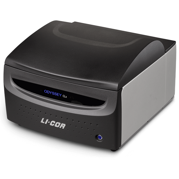
Get clearer answers with accurate, reproducible Western blots and many other assays.
Capability:
Measure concentration and purity of DNA, RNA or protein samples.
Do more with consistent, reproducible digital imaging technology that has propelled the Odyssey imager family to over 10,000 peer-reviewed publications. See the benefits of stable NIR fluorescence and detect multiple targets in the same lane. Get the most accurate results with over 6 logs of linear dynamic range, so you can make more discoveries.
Core Facility: Human Embryonic Stem Cell Core
SAMPLE SHEARING & ISOLATION
Covaris E220 Focused Ultra Sonication - Covaris
Instrument:
Covaris E220
Capability:DNA Fragmentation, Chromatin Shearing
Core Facility: Stem Cell Genomics Core
SEQUENCING
MiSeq – illumina
Instrument:
Capability:
Highly multiplexed amplicon sequencing, Targeted Resequencing, Small Genome Sequencing, De Novo sequencing, CHiP Seq, Small RNA Sequencing, Library QC for HiSeq
*MiSeq not recommended for deep sequencing or sequencing of large genomes. For deep sequencing on the HiSeq, please contact IGM
Core Facility: Stem Cell Genomics Core
SINGLE-CELL LIBRARY PREPARATION
Chromium X - 10x Genomics

Capability:
Compact, sleek, efficient.
Chromium X makes single-cell analysis easy, reliable, and scalable thanks to its proprietary Next GEM technology. The key to this technology is the ability to generate hundreds of thousands of single-cell partitions, each containing an identifying barcode for downstream analysis. Combined with an innovative reagent delivery system and turnkey software analysis tools, we enable the discovery of previously inaccessible biological information. In addition, the Chromium X features fully integrated, automated system support in the 10x Genomics Cloud, complete with temperature control and improved access to support.
Core Facility: Stem Cell Genomics Core
C1 Single Cell Auto Prep – Fluidigm
Fluidigm C1
Capability:Gene Expression, Single-Cell Gene Expression, Single-Cell mRNA Sequencing Aapplication, provides cDNA from single cell, utilizes NEXTera XT kit for subsequent manual library prep
Core Facility: Stem Cell Genomics Core
THERMOCYCLER
ProFlex 3x32-well PCR System - Thermo Fisher Scientific
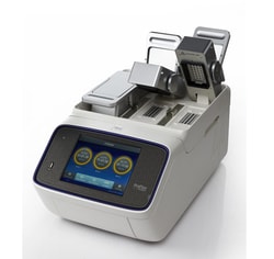
Capabilities:
The ProFlex™ 3x32-Well PCR System is the latest in our line of Applied Biosystems® thermal cyclers. The ProFlex™ system combines the reliability and performance you’ve come to expect from Applied Biosystems® instruments with the flexible configuration and control features that adapt to your research needs.
Request a demo.
The ProFlex™ 3x32-Well PCR System allows you to:
• Run three experiments at once with the 3x32-well, 0.2-mL block
• Interchange the 3x32-well block with another block type if your throughput needs change
• Access the system remotely (and conveniently) through a mobile app
• Program the instrument in seconds with a simple-to-use touchscreen interface
• Simulate your old instrument with Thermal Simulation modes
• Optimize your PCR quickly and efficiently with better-than-gradient VeriFlex™ blocks
Core Facility: Stem Cell Genomics Core
Thermal Cycler C1000 Touch 96w - Bio-Rad
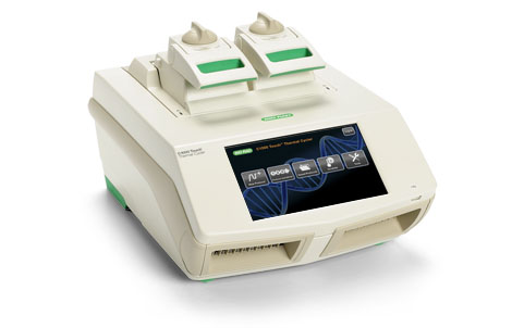
Thermal Cycler C1000 Touch 96w
Capabilities:
The C1000 Touch thermal cycler offers superior performance and a large color touch screen for easy programming. This fully modular platform is able to accommodate different throughput needs with easily interchangeable reaction modules that swap in seconds without tools. Each PCR module has a fully adjustable heated lid that supports a wide range of vessels and sealers, including low-profile and standard-height PCR plates.
Features and Benefits
- Quick and easy protocol programming — 2 programming options, including the Protocol Autowriter, which automatically generates customized standard, fast, and ultrafast protocols
- Easy protocol editing — the large color touch screen and redesigned navigation make editing and running protocols easy
- Flexible platform — change throughput capability by linking up to 3 additional S1000 thermal cyclers or adding PC control for up to 32 cyclers
- Advanced file management — USB flash drive compatibility allows universal protocol transfer and unlimited data storage
- Thermal gradient — quickly and easily identify optimal annealing temperatures by using the programmable temperature gradient
Applications and Uses
- Amplification/PCR
- Cloning
- Cycle sequencing
- Gene expression studies
- Mutagenesis
The C1000 Touch thermal cycler can be used with six interchangeable reaction modules, including:
- A gradient-enabled 96-well fast module
- A gradient-enabled 96–deep well module
- A gradient-enabled dual 48/48-well fast module that allows 2 independently controlled protocols to be run side by side in a single bay
- A gradient-enabled 384-well module for high throughput
- 2 modules for real-time PCR detection
Core Facility: Stem Cell Genomics Core
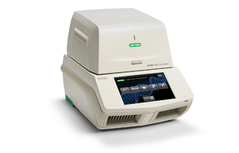
BioRad CFX 384
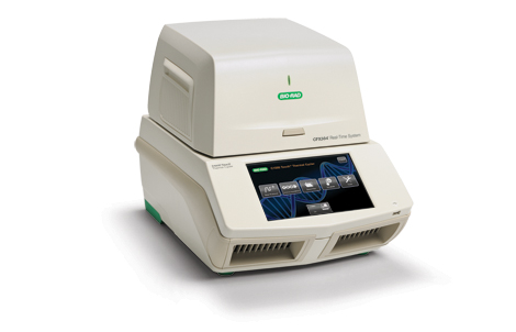
Thermal Cycler C1000 Touch 96w
Capabilities:
The CFX384 Touch Real-Time PCR Detection System is a powerful and precise real-time PCR instrument in a 384-well format, for researchers who require both ease of use and rapid, high-throughput data acquisition. The CFX384 Touch System utilizes accurate temperature control and sensitive, 4-color detection optics to deliver reliable and repeatable qPCR results for singleplex or multiplex reactions.
Quickly set up runs and monitor amplification traces in real time on the integrated LCD touch screen, or use CFX Maestro Software to easily and intuitively design your experiment and analyze results from a connected computer. With up to four-target detection, unsurpassed thermal cycler performance, unrivaled stand-alone functionality, and powerful software, the CFX384 Touch System was designed to advance your qPCR.
Key Features and Benefits
With the CFX384 Touch Real-Time PCR Detection System you can:
- Obtain precise quantification and multiplex target discrimination – optical system uses long-lasting solid-state technology with 5 filtered LEDs and 5 filtered photodiodes
- Configure the system to fit your laboratory needs – optimized design lets you run without a computer, run up to 4 systems from 1 computer, or integrate with the CFX Automation System II for higher throughput
- Confirm system performance and data reliability – internal system qualification and system test software, automated data quality control, and various validation tool services are available
- Obtain accurate results with volumes as low as 3 µl – minimize sample and reagent usage
- Easily integrate with your laboratory information management system (LIMS) – integrate with any LIMS using built-in LIMS file management
- Combine the CFX384 Touch System with good laboratory practice standards – use CFX Maestro Software, Security Edition, which complies with U.S. FDA 21 CFR Part 11 regulations, for data collection and analysis
Core Facility: Human Embryonic Stem Cell Core
BioRad CFX 96
Capabilities:
The CFX96 Touch System is a powerful, precise, and flexible real-time PCR detection system. This six-channel (five colors and one FRET channel) real-time PCR instrument combines advanced optical technology with precise temperature control to deliver sensitive, reliable detection for singlexplex or multiplex reactions.
Quickly set up runs and monitor amplification traces in real time on the integrated LCD touch screen, or use the included CFX Maestro Software to easily and intuitively design your experiment and analyze results from a connected computer. With up to five-target detection, unsurpassed thermal cycler performance, unrivaled stand-alone functionality, and powerful yet easy-to-use software, the CFX96 Touch System is designed to advance your qPCR.
Key Features and Benefits
With the CFX96 Touch Real-Time PCR Detection System you can:
- Set up your system quickly — easy installation and factory-calibrated optics
- Minimize sample and reagent use — up to 5-target multiplexing with sample volumes as low as 10 µl
- Optimize reactions in a single run — thermal gradient feature
- Analyze data faster — visualize all run data at once and export only the data you need in the format you want
- Use advanced data analysis tools — normalized gene expression using CFX Maestro Software
- Configure the system to fit your needs — run without a computer, run up to 4 systems from 1 computer, or integrate with the CFX Automation System II for higher throughput
- Combine the CFX96 Touch System with good laboratory practice standards — use CFX Maestro Software, Security Edition for data collection and analysis to simplify compliance with U.S. FDA 21 CFR Part 11 regulations
Flexible CFX System Configurations Meet Your Throughput Needs
The CFX Automation System II works with all CFX Real-Time PCR Detection Systems to enable walk-away, high-throughput qPCR operation. Integrate this robotic plate handler with one or two CFX Systems to maximize throughput while maintaining a compact footprint.
Core Facility: Human Embryonic Stem Cell Core
ULTRACENTRIFUGE
Optima L-80 XP Ultracentrifuge 1
Optima L-80 XP Ultracentrifuge 1
Capability:
The ultracentrifuge is a centrifuge optimized for spinning a rotor at very high speeds. The L-80 XP is capable of generating 602,000 x g at speeds of up to 80,000 rpm. The L-80 XP, along with a broad range of rotors, tubes, and supplies shortens run times for a wide range of applications. The L-80 XP’s special design eliminates the need for chlorofluorocarbons or other ozone-depleting coolants for drive and chamber cooling.
Instrument
Thermal Cycler C1000 Touch 96w
Capabilities:
The C1000 Touch thermal cycler offers superior performance and a large color touch screen for easy programming. This fully modular platform is able to accommodate different throughput needs with easily interchangeable reaction modules that swap in seconds without tools. Each PCR module has a fully adjustable heated lid that supports a wide range of vessels and sealers, including low-profile and standard-height PCR plates.
Features and Benefits
- Quick and easy protocol programming — 2 programming options, including the Protocol Autowriter, which automatically generates customized standard, fast, and ultrafast protocols
- Easy protocol editing — the large color touch screen and redesigned navigation make editing and running protocols easy
- Flexible platform — change throughput capability by linking up to 3 additional S1000 thermal cyclers or adding PC control for up to 32 cyclers
- Advanced file management — USB flash drive compatibility allows universal protocol transfer and unlimited data storage
- Thermal gradient — quickly and easily identify optimal annealing temperatures by using the programmable temperature gradient
Applications and Uses
- Amplification/PCR
- Cloning
- Cycle sequencing
- Gene expression studies
- Mutagenesis
The C1000 Touch thermal cycler can be used with six interchangeable reaction modules, including:
- A gradient-enabled 96-well fast module
- A gradient-enabled 96–deep well module
- A gradient-enabled dual 48/48-well fast module that allows 2 independently controlled protocols to be run side by side in a single bay
- A gradient-enabled 384-well module for high throughput
- 2 modules for real-time PCR detection
Core Facility: Human Embryonic Stem Cell Core
Optima L-80 XP Ultracentrifuge 2
Optima L-80 XP Ultracentrifuge 2
Capability:
The ultracentrifuge is a centrifuge optimized for spinning a rotor at very high speeds. The L-80 XP is capable of generating 602,000 x g at speeds of up to 80,000 rpm. The L-80 XP, along with a broad range of rotors, tubes, and supplies shortens run times for a wide range of applications. The L-80 XP’s special design eliminates the need for chlorofluorocarbons or other ozone-depleting coolants for drive and chamber cooling.
Core Facility: Human Embryonic Stem Cell Core